
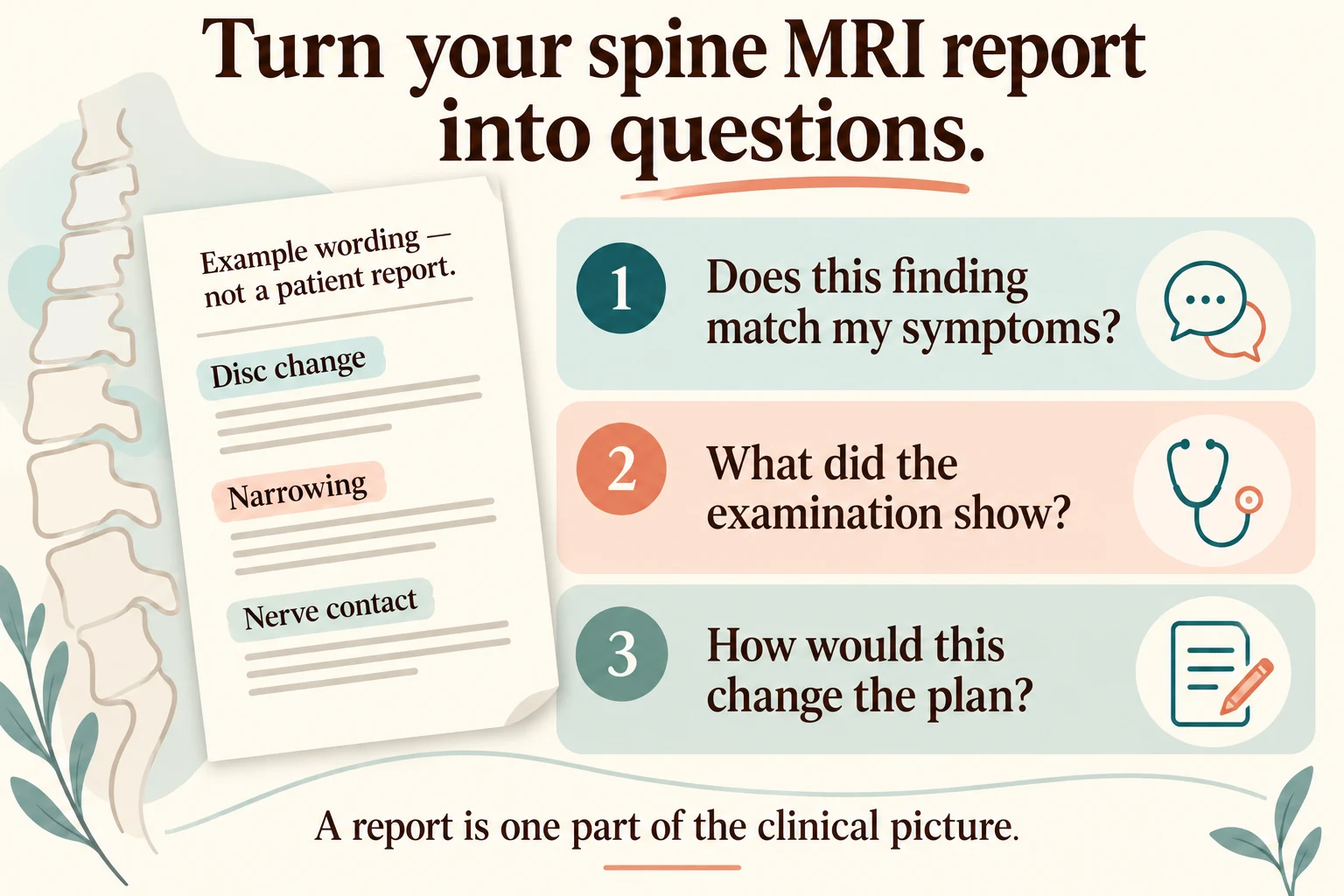
Original educational illustration. A clearly fictional report with disc change, narrowing and nerve-contact wording sits beside three consultation questions.
Ask whether a finding matches your symptoms, what the examination showed, and how it would change the plan. The example is not a patient report or a diagnostic image.
Illustration references: Reference 1 · Reference 2 · Reference 3
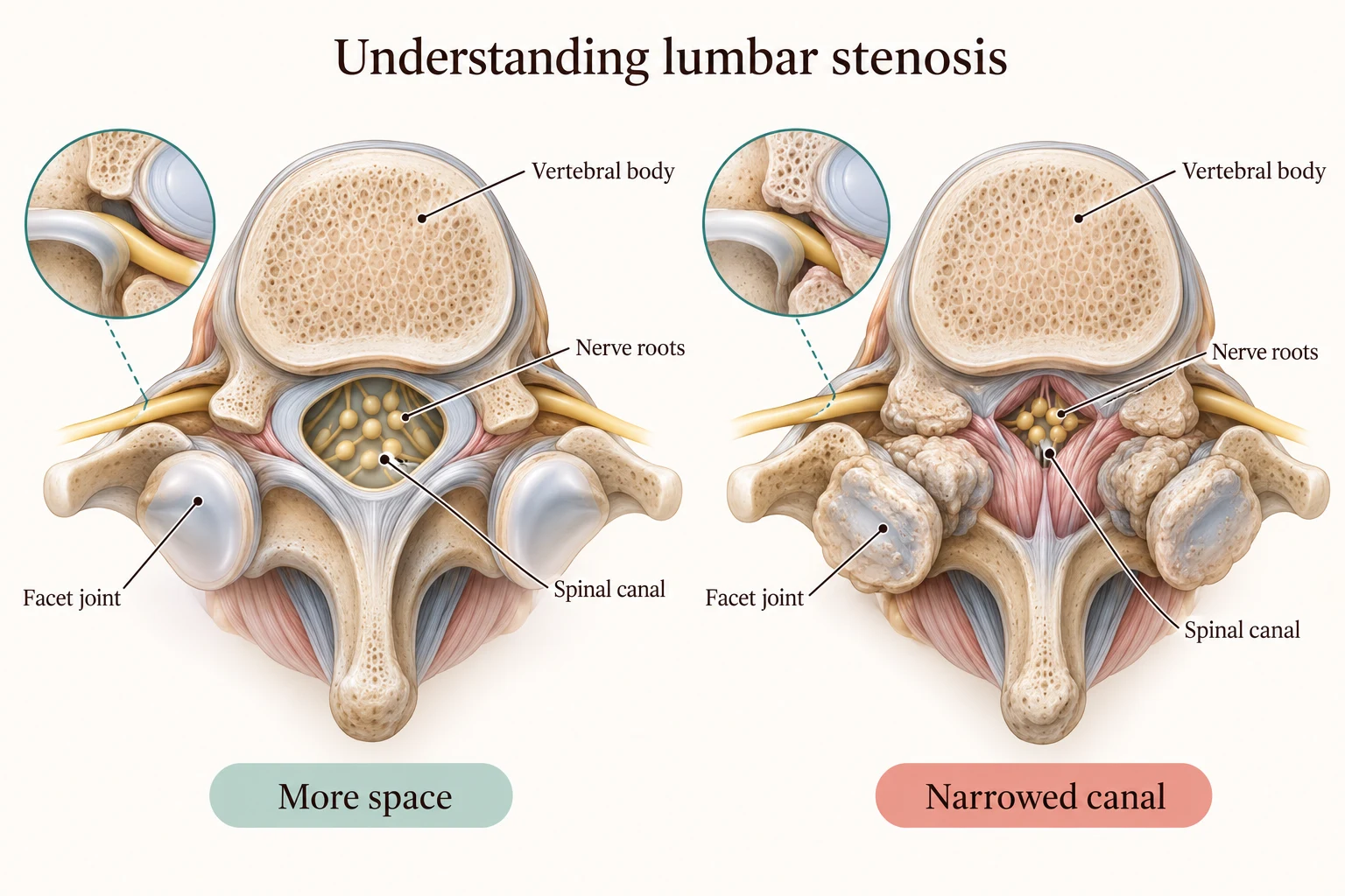
Original educational illustration. Two schematic cross-sections compare more space and a narrowed lumbar spinal canal.
The lower lumbar canal contains nerve roots. A picture of narrowing does not establish the cause of a person's symptoms; the examination and imaging need to be considered together.
Illustration references: Reference 1 · Reference 2 · Reference 3
1. What question was the scan meant to answer?
Spine MRI uses a magnetic field and radio waves to create detailed images. A radiologist interprets them and sends a report to the referring clinician. The report is one part of a clinical assessment, rather than a treatment plan you are expected to interpret alone.
Ask why the MRI was ordered and whether it answered the original question. Was the concern a nerve problem, a change after surgery, or something else? That context helps you separate useful findings from unfamiliar phrases that do not affect the decision. If the report appears in a portal before your appointment, mark your questions and confirm who will review the results with you and when.
Sources: Spine MRI
2. Which finding matches my symptoms and examination?
HSS emphasizes matching cervical imaging with the patient’s symptoms and examination because disc changes and bone spurs do not always cause pain. The general question is useful whenever you discuss a spine report: what clinical evidence makes this particular finding relevant?
Bring a brief description of where symptoms occur, which side is affected, how they travel, and what function has changed. Ask the clinician to show the level and structure thought to explain that pattern. If the report highlights a different side from the one that bothers you, ask how the mismatch is interpreted. If several levels look abnormal, ask which one or two findings actually guide the next step.
Sources: Cervical Radiculopathy
3. Can you translate the location and terminology into plain language?
Ask what the unfamiliar term refers to: a disc, joint, the central canal, a nerve exit opening, or another structure. Have the clinician identify whether a discussion concerns a nerve root, spinal cord, or some other problem. The relevant anatomy is easier to understand when it is shown on your own images.
You do not need to memorize every abbreviation or measurement. Ask for the short explanation you could give a family member. If a report uses words such as narrowing, bulge, or degeneration, ask what each contributes to the working diagnosis in your case. Keep a note of the terms that change management and the terms that are background information.
Sources: Spine MRI
4. What does the severity wording mean for the decision?
A report may describe a finding as mild, moderate, or severe. Ask the treating clinician how that wording relates to your examination and function. A severity adjective does not explain by itself whether an operation is needed or which symptom a proposed treatment would address.
If the report sounds alarming but the recommendation is observation or rehabilitation, ask why. If the images seem modest but symptoms are very limiting, ask what else should be assessed. Be clear about the activity you most want to regain and whether the finding plausibly explains that limitation. The purpose of the discussion is a meaningful interpretation, not reassurance based solely on the wording of the scan.
Sources: Cervical Radiculopathy
5. What is still uncertain, and would another test resolve it?
ACR and RSNA patient guidance describes how the usefulness of low-back imaging depends on the clinical situation. Ask what a proposed repeat MRI, contrast study, CT, X-ray, or nerve test would add. The important follow-up is how each possible result could change care.
If you already have recent studies, ask whether the team needs the actual files or a secure transfer for comparison. Do not arrange duplicate testing before checking what is needed. If contrast or another MRI is planned, tell the imaging team about implants, prior procedures, and relevant medical concerns so they can give appropriate safety instructions. A test deserves a clinical question, not just a place on a checklist.
Sources: Low Back Pain: Appropriateness Criteria · Spine MRI
6. Does the finding change treatment or the urgency of assessment?
Ask whether the next step is further clinical assessment, nonsurgical treatment, monitoring, or a surgical discussion, and why. If a procedure is proposed, ask which image finding supports the target and what symptom it should address. If no immediate change is needed, agree on what will be monitored.
Do not wait for a routine report visit if you develop new loss of bowel or bladder control, numbness around the groin or saddle area, or severe new weakness. These need urgent assessment. For other changes, ask the team which contact route to use. A reassuring interpretation of an earlier MRI does not replace assessment of a new symptom.
Sources: Lumbar Spinal Stenosis
7. What should I bring to the next conversation?
Bring the report, the actual images if requested, earlier studies, and a short treatment timeline. Include medicines, therapy, injections, and prior operations, with what changed after each. Choose the two questions you most want answered rather than trying to discuss every phrase in the report at once.
Before the visit ends, request a summary: which finding matters, what it may explain, what remains uncertain, and the next step. If another clinician is involved, ask how the assessment will be shared. Keep that summary with your records. It gives a future appointment a clearer starting point and helps you discuss the plan with the people supporting you.
Our spine and pain clinicians assess imaging alongside symptoms and examination. Dr. Amandeep Bhalla, Dr. Brandon Ortega, and Dr. Evish Kamrava bring different perspectives to spine concerns. Explore their profiles and bring the question you most want resolved. We can use the report as a starting point for a conversation about your own care.
Sources: Spine MRI
Make the most of your visit
Questions to bring to your consultation
Choose the questions that matter most to you. Bring them along, and ask us to explain anything that is unclear.
- What question was my MRI ordered to answer?
- Which finding matches my symptoms, side, and examination?
- Can you show me the level and structure in plain language?
- How does the report’s severity wording affect the decision?
- What remains unexplained, and would another test change care?
- Does the finding change treatment or the urgency of follow-up?
- Which new symptoms require assessment before the scheduled visit?
- Can you summarize the relevant findings and next step for me?
Sources & further reading
- Spine MRIACR / RSNA RadiologyInfo
- Cervical RadiculopathyHospital for Special Surgery
- Low Back Pain: Appropriateness CriteriaAmerican College of Radiology and Radiological Society of North America
- Lumbar Spinal StenosisJohns Hopkins Medicine
We use these medical sources to help explain your options and prepare you for a conversation with our team. Our guides feature our own clinicians; patient reviews describe individual experiences.








 (310) 595-1030